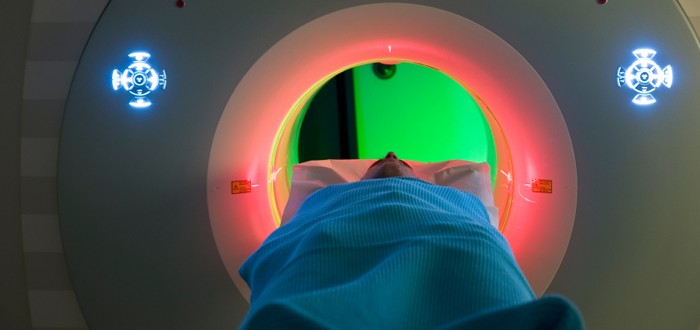
Researchers in England have concerns that using just a CT scan to identify malignant mesothelioma may miss pleural malignancies and lead to incorrect diagnoses.
A “significant proportion” of patients examined for suspected mesothelioma with the aid of nothing more than a CT scanner can expect to be told they do not have the disease, when actually they do.
Researchers from the Respiratory Trials Unit of Oxford University in England report that CT scans of the chest have a tendency to produce falsely negative findings.
Thinking their patients are mesothelioma-free, the doctors order no treatments. This causes tragic delay in the start of any counterattack that could be launched against the cancer.
Therefore, mesothelioma doctors should reconsider the use of CT images alone to give a mesothelioma diagnosis, the researchers recommend.
CT Still a Useful Mesothelioma Diagnosis Tool
The researchers note that there is an ongoing debate among mesothelioma specialists as to which clinical tools and exam pathways deliver the most reliable diagnoses.
Writing in a recent issue of the journal Thorax, the researchers explain why they believe a CT scanner should not be the sole tool used.
They begin by describing a retrospective review they conducted of mesothelioma biopsy cases at two British hospitals over a five-year period ending in 2013.
In the course of that review, the researchers turned up 370 patients who were biopsied and had it confirmed that they were afflicted with pleural tumors.
Among these patients, the tumors in 211 were revealed to be malignant. But a CT scan performed beforehand on each of the 211 detected malignancy in just 144 of them.
The researchers calculated that this meant the CT scans had a sensitivity score of about 65 percent for finding malignant pleural tumors.
The biopsies of the remaining 159 patients revealed the tumors to be benign. In this group, the pre-biopsy CT scan did better — it correctly recognized the growths to be benign in 124 of them.
The researchers calculated this to mean the CT scans had a sensitivity score of roughly 80 percent for identifying benign tumors.
“The aim of this study was to assess the sensitivity and specificity of CT in detecting pleural malignancy prior to definitive histology,” they wrote.
CT Use for Mesothelioma Diagnosis Is Up
This research is important because the use of CT scans to help detect mesothelioma has greatly increased during the past 20 years.
Mesothelioma doctors find CT more useful than conventional chest X-rays. For one thing, CT offers much higher image resolution, so tumors really stand out and are less likely to be overlooked.
For another thing, CT shows only those anatomic structures that the doctors really want to study.
Chest X-rays, on the other hand, show structures in front of and behind the area of actual interest. That tends to obscure the view, making it harder to see if cancer is there.
CT works by using X-rays, just like any conventional X-ray machine. But CT uses them in a very different way.
With conventional X-ray, the radiation beamed into the patient comes from a stationary source. With CT, the X-ray source rotates completely around the patient.
In both conventional X-ray and CT, the radiation that passes through the patient’s body is picked up on the other side by a detector device.
An X-ray machine’s detector sends what it captures to a sheet of film. A CT scanner’s detector sends what it captures to a computer that generates a richly detailed image for viewing on a monitor.
Because the CT’s X-rays were beamed in a circle around the patient, doctors can look at the image in 3-D and even perform a virtual “walk-around” of the tumor and view it from every conceivable angle.
For these reasons, CT is a valuable tool in the fight against mesothelioma.
CT might or might not have trouble telling a malignant tumor from a benign one. But it has almost no trouble seeing a growth that’s not supposed to be there.
CT sees exactly what that tumor’s size is today and will show exactly how much bigger it has become the next time the growth is scanned.

