One of the first mesothelioma diagnosis tests you’ll be given when your doctor suspects you’ve got the deadly cancer is thoracentesis.
This test cytologically analyzes a sample of the pleural fluid in your lungs to see whether it contains telltale traces of mesothelioma.
Unfortunately, the mesothelioma diagnostic yield of thoracentesis isn’t usually as bountiful as doctors would like, so the test must be repeated or you must be given other kinds of tests — an invasive biopsy, for instance.
Doctors have been understandably keen about improving the diagnostic yield of thoracentesis testing. They need more reliable determinations of if those symptoms you’ve been experiencing are actually signs of mesothelioma. The doctors need to determine if mesothelioma cells are growing on the lining of your lungs and developing into mesothelioma tumors.
One such improvement is the cell-block method of pleural fluid cytological analysis.
Researchers recently unveiled findings that suggest the cell-block method is better than the traditional method when it comes to obtaining a greater diagnostic yield.
Study Focused on More than Mesothelioma
The researchers, affiliated with Selcuk University’ s medical school in Konya, Turkey, published findings in the Asian Pacific Journal of Cancer Prevention. They arrived at their conclusions about the cell-block method by studying 194 patients with exudative pleural effusions.
These were not only mesothelioma patients, however. They were patients with lung cancer generally — only a couple of mesothelioma cases were to be found among them.
From each patient, the researchers extracted 10 ml of fresh pleural fluid and then split up the collected samples into two batches.
The first was earmarked for cytological analysis the traditional way. The second was slated to be analyzed using the cell-block technique.
The samples in each group were further divided into the categories of benign, malignant and status undetermined.
The researchers said they performed histopathological diagnosis once they had harvested sufficient fluid samples containing sufficient numbers of cancer cells.
Mesothelioma Cases Diagnosed in Group
Using the traditional method, they determined that 154 of the patients had benign conditions. But, using the cell-block method, they found only 147 patients with benign conditions.
This means that seven patients whose doctors relied on the traditional method would have been given a clean bill of health when in fact they were harboring malignancies.
The researchers reported that the cell-block method — in addition to being better at detecting the presence of cancer – also detected cancer with specificity.
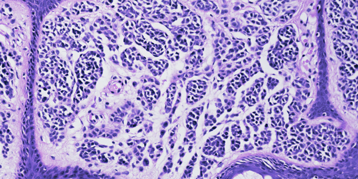
Using the cell-block method, the researchers were able to discern that 12 of the patients with malignancies had metastatic cancer, four had squamous cell carcinoma, 18 had adenocarcinoma, five had large-cell carcinoma, two had mesothelioma, three had small cell carcinoma, and three had lymphoma.
“Our study confirmed that the cell-block method increases the diagnostic yield with exudative pleural effusions accompanying lung cancer,” the researchers wrote.
The study was entitled “Importance of the Cell Block Technique in Diagnosing Patients with Non-Small Cell Carcinoma Accompanied by Pleural Effusion.”
This research builds on and confirms a 2013 study, The Cell Block Method Increases the Diagnostic Yield in Exudative Pleural Effusions Accompanying Lung Cancer,” published in the Turkish Journal of Pathology and involving a much smaller number of patients. That team of researchers was from the Atatürk Chest Diseases and Chest Surgery Training and Research Hospital in Ankara, Turkey.