Endoscopic ultrasound-guided tissue acquisition is an easier and more reliable way to diagnose peritoneal mesothelioma than are invasive procedures such as laparoscopy, say researchers in Japan.
In a letter to the editor of the journal Endoscopic Ultrasound, the researchers described their success at acquiring suspect abdominal cavity tissues by use of endoscopic ultrasound.
This was not an actual clinical study because the tissue-acquisition technique was tried on only one patient. Even so, the researchers were quite excited about the results.
They hoped that sharing their results might inspire others to investigate endoscopic ultrasound-guided acquisition of peritoneal tissues for diagnosis.
Tissue Mass Revealed as Peritoneal Mesothelioma
The letter was written by several researchers from the Department of Gastroenterology in the Graduate School of Medicine at The University of Tokyo, Japan.
They explained how they used endoscopic ultrasound-guided tissue acquisition on a patient to identify a tumor mass as peritoneal mesothelioma.
The patient was a 73-year-old man with a history of sigmoid colon cancer. The researchers first saw him in 2010. At that time he presented with an abdominal mass requiring identification.
Starting with conventional diagnostic procedures, the researchers determined the patient had a 3 cm, ill-defined, soft tissue mass surrounding the second portion of his duodenum.
Further diagnostic procedures revealed a hypoechoic mass infiltrating both the liver and duodenum.
At this point, the researchers switched to endoscopic ultrasound to perform a through-the-needle biopsy and a fine-needle aspiration.
The needle used in the tissue acquisition and aspiration was a 19-G preloaded with 0.75-mm miniature biopsy forceps.
Under endoscopic ultrasound guidance, the needle was brought into contact with the lesion. It took just one pass following puncture of the lesion to aspirate sample tissue back up through the tube.
It was simple and quick, and it revealed malignant pleural mesothelioma.
About Endoscopic Ultrasound Procedures
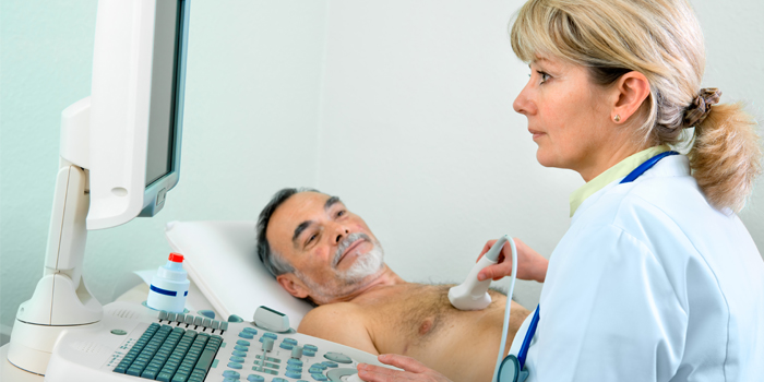
As the name suggests, endoscopic ultrasound requires the use of an endoscope. This is a thin tube designed to be inserted through a bodily opening and fed into a hollow organ, such as the chest or abdomen.
The endoscope is used by the doctor to get an up-close inside view of those hollow organs.
The endoscope can also be used by the doctor to poke at, cut into or grab tissues within the organs. This is accomplished by attaching operable special tips to the endoscope.
Ultrasound is an imaging technology that bounces sound waves off the internal organs to render a picture of them. It operates on the same basic principle as submarine sonar.
What the researchers from Japan did was harness ultrasound imaging to show them exactly where the tumor mass was growing inside the patient’s abdomen.
That enabled them to precisely position the endoscope above the tumor. As a result, the tissues they retrieved were in fact those from the tumor and nowhere else.
The quality of the ultrasound image produced for a purpose like this depends on the sound wave frequency that is used. The rule of thumb is the higher the frequency, the better the picture.
The researchers note that they are not the first to use endoscopic ultrasound for diagnosing mesothelioma. However, earlier users have employed it almost exclusively in pleural mesothelioma cases.